


Pterosoma
Pterosoma planum
Roger R. Seapy
Introduction
Pterosoma is a monotypic genus represented by the Indo-Pacific species P. planum. The transparent trunk, with whitish minute dish-shaped inclusions and surface tubercles, is expanded as an oblong disk that is flattened dorso-ventrally. The trunk terminates in a tapering tail. The visceral nucleus arises from a short, mobile stalk and is covered by a flattened shell. The shell is divided medially into two lateral halves by a low carina, homologous with the keel in Carinaria. The carina runs the length of the shell, beginning at the posterior end abutting the protoconch. While swimming, the visceral nucleus is raised dorsally, exposing the gills to inflowing water, but if disturbed it is retracted into a depression on the trunk surface. The proboscis is moderately short, extending ventrally beneath the anterior margin of the trunk.
Brief Diagnosis
Carinariid heteropods with:
- Trunk expanded, forming an oblong disk that is flattened dorso-ventrally
- Visceral nucleus arises from short stalk and is covered by a flattened shell
- Tapering tail extends posteriorly from trunk
- Proboscis extends ventrally from beneath anterior margin of trunk
Characteristics
- Body morphology
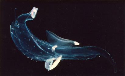
- Trunk greatly expanded laterally, forming an oblong disk that is flattened dorso-ventrally Click on an image to view larger version & data in a new window
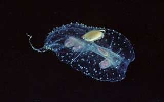
Figure. Dorsal view of free-swimming Pterosoma planum. ©
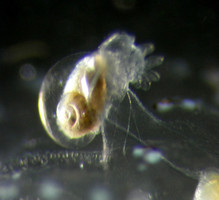
- Trunk highly transparent, covered on the surface with whitish tubercles and embedded with minute whitish, disk-shaped inclusions Click on an image to view larger version & data in a new window
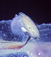
Figure. Dorso-lateral view of Pterosoma planum, with elevated visceral nucleus and exposed gills. Low whitish tubercles and imbedded minute, disk-shaped inclusions are seen on surface of the trunk to the left and below the gills. ©
- Visceral nucleus dorso-ventrally compressed, arising from a flexible short stalk (see above photo)
- Fin sucker present in both males and females
- Tail extends posteriorly from beneath the trunk, tapering to a narrow terminal section (see first photo above)
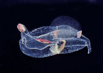
- In young, post-metamorphic individuals, the expanded trunk disk (about 5 mm in length in the specimen below) is well developed and is fringed by about two dozen, prominent white tubercles. Numerous small white inclusions are also evident in the disk. Also, at this stage the visceral nucleus stalk is prominent and the adult shell (teleoconch) is just beginning to form (see second image below)
Click on an image to view larger version & data in a new window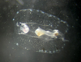
Click on an image to view larger version & data in a new windowFigure. Juvenile specimen of Pterosoma planum. Left: Ventral view; note the large white tubercles on the fringe of the trunk disk, the multiple small white inclusions in the disk, the single left tentacle anterior to the left eye, and the yellowish-tan bolus of food in the esophagus (central part of body). Right: Antero-lateral view of visceral nucleus and shell. At this stage of development the adult teleoconch is just beginning to form. © Allan Connell
- Trunk greatly expanded laterally, forming an oblong disk that is flattened dorso-ventrally
- Shell
- Adult shell dorso-ventrally compressed, covering the visceral nucleus and gills (see first image below)
- Shell divided medially (in the anterior-posterior axis) by a low carina whose height increases somewhat with shell growth
- Coiled protoconch (larval shell) at posterior end of teleoconch (adult shell)Click on an image to view larger version & data in a new window
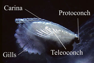
Figure. Shell of Pterosoma planum viewed from the right side. Protoconch at posterior end of shell. Shell divided medially by low carina, which increases slowly height with shell growth. ©
- Larval shell globular. Right side with smooth surface except for low radiating ridges on second whorl (first image below). Spire low and aperture broadly oval and indented medially (second image below). Left side of shell with open umbilicus, having about 12 radiating striae on wall (third image below)
Click on an image to view larger version & data in a new window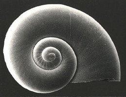
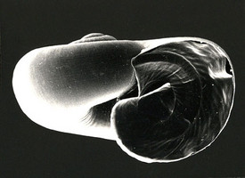
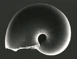
Figure. Larval shell of Pterosoma planum; views of right side, aperture and left side, respectively. Shell diameter = 0.70 mm (right and middle images) and 0.65 mm (right image). ©
- Adult shell dorso-ventrally compressed, covering the visceral nucleus and gills (see first image below)
- Swimming fin well-developed, with a large fin sucker that is located dorsally on the posterior margin of the fin (see title illustration). Fin sucker present in both sexes
- Eyes and tentacles
- Eyes broadly triangular in dorsal view (see photograph below)
- Left tentacle large and right one very reduced (see below and title illustration)Click on an image to view larger version & data in a new window
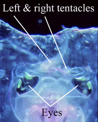
Figure. Dorsal view of head region in Pterosoma planum. Right and left tentacles (labelled) are somewhat out of focus. ©
Behavior
Observations of free-swimming Pterosoma planum in aquaria aboard ship (Seapy, unpublished) revealed that the stalked visceral nucleus is normally elevated dorsally, exposing the gills and the visceral nucleus to inflowing water (see photographs under 1.a above). However, when individuals were prodded with a blunt probe, the visceral nucleus (covered dorsally by the flattened shell) was rapidly retracted into a longitudinal groove on the dorsal surface of the trunk. Hypothetically, the act of prodding an individual simulates disturbance by a predator, and the vulnerable soft tissues of the gills and visceral nucleus are protected beneath the shell.
In the book on holoplanktonic gastropod snails by Lalli and Gilmer (1989:32), the in-situ field observations of swimming Pterosoma planum by Gilmer were described: "Pterosoma has large opalescent spots covering the central body in both sexes; these spots continually change in brilliance and pattern as the animals swim." While the "central body" undoubtedly refers to the trunk, the nature of the "opalescent spots" is unclear since the only structures clearly visible on the trunk surface in net-captured specimens (Seapy, pers. obs.) are the surface tubercles described above under 1.b. Conceivably, the "opalescent spots" and the behavior observed by Gilmer may be limited to animals in-situ. Despite observing active and apparently healthy specimens swimming freely in aquaria aboard ship for extended periods of time (Seapy, pers. obs.), the observations of Gilmer were never seen.
References
Lalli, C. M. and R. W. Gilmer. 1989. Pelagic snails. The biology of holoplanktonic gastropod snails. Stanford: Stanford University Press. 259 pp.
Richter, G. and R. R. Seapy. 1999. Heteropoda, pp. 621-647. In: D. Boltovskoy (ed.), South Atlantic Zooplankton. Leiden: Backhuys Publishers.
Seapy, R. R. and C. Thiriot-Quiévreux. 1994. Veliger larvae of Carinariidae (Mollusca: Heteropoda) from Hawaiian waters. Veliger 37: 336-343.
Title Illustrations

| Scientific Name | Pterosoma planum |
|---|---|
| Location | Florida Current |
| Specimen Condition | Live Specimen |
| Sex | Male |
| Life Cycle Stage | adult |
| View | right side |
| Copyright | © Ronald Gilmer |

| Scientific Name | Pterosoma planum |
|---|---|
| Location | Hawaiian waters |
| Specimen Condition | Live Specimen |
| Sex | Male |
| Life Cycle Stage | adult |
| View | ventral |
| Image Use |
 This media file is licensed under the Creative Commons Attribution-NonCommercial License - Version 3.0. This media file is licensed under the Creative Commons Attribution-NonCommercial License - Version 3.0.
|
| Copyright |
©

|
About This Page
California State University, Fullerton, California, USA
Correspondence regarding this page should be directed to Roger R. Seapy at
Page copyright © 2007
 Page: Tree of Life
Pterosoma . Pterosoma planum .
Authored by
Roger R. Seapy.
The TEXT of this page is licensed under the
Creative Commons Attribution License - Version 3.0. Note that images and other media
featured on this page are each governed by their own license, and they may or may not be available
for reuse. Click on an image or a media link to access the media data window, which provides the
relevant licensing information. For the general terms and conditions of ToL material reuse and
redistribution, please see the Tree of Life Copyright
Policies.
Page: Tree of Life
Pterosoma . Pterosoma planum .
Authored by
Roger R. Seapy.
The TEXT of this page is licensed under the
Creative Commons Attribution License - Version 3.0. Note that images and other media
featured on this page are each governed by their own license, and they may or may not be available
for reuse. Click on an image or a media link to access the media data window, which provides the
relevant licensing information. For the general terms and conditions of ToL material reuse and
redistribution, please see the Tree of Life Copyright
Policies.
- First online 10 December 2007
- Content changed 15 September 2008
Citing this page:
Seapy, Roger R. 2008. Pterosoma . Pterosoma planum . Version 15 September 2008. http://tolweb.org/Pterosoma_planum/28743/2008.09.15 in The Tree of Life Web Project, http://tolweb.org/


