
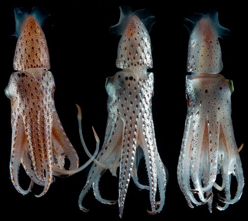
Figure. Ventral, ventral and dorsal views of H. bonnellii, showing two color phases. Photographed alive in a shipboard aquarium by
- Photophores: Compound photophores (Type 1b)
- Basal Arm IV Row with 3 photophores and separated medially by 1 photophore from anterior photophore of Midline Series.
- Midline Series with 3 photophores.
- Right Eyelid Series with 17 photophores (rarely 16 or 18: Voss, et al.,1998).
- Left Eyelid Series with 6 photophores.
- 2° Right Eyelid Series with 8 photophores.
- 2° Left Eyelid Series with 7 photophores.
- Ventral Matrix with 36 photophores including an asymmetrically placed dangling, rogue photophore (colored red in illustration), and 9 longitudinal (blue) series. Attachment to 2° Eyelid Series at 2° eyelid photophores no. 2 either side.
- Ventral Matrix photophores do not align in transverse rows.
- Right Basal Series with 2 photophores.
- Left Basal Series with 3 photophores, medial two have very large bases (see below).
- Basal Row with 7 photopores and one sawtooth (note alignment of left sawtooth photophores with the "dangling" (red) photophore.
- Right Accessory Series with 2 photophores.
- Left Accessory Series with 3 photophores.
Click on an image to view larger version & data in a new window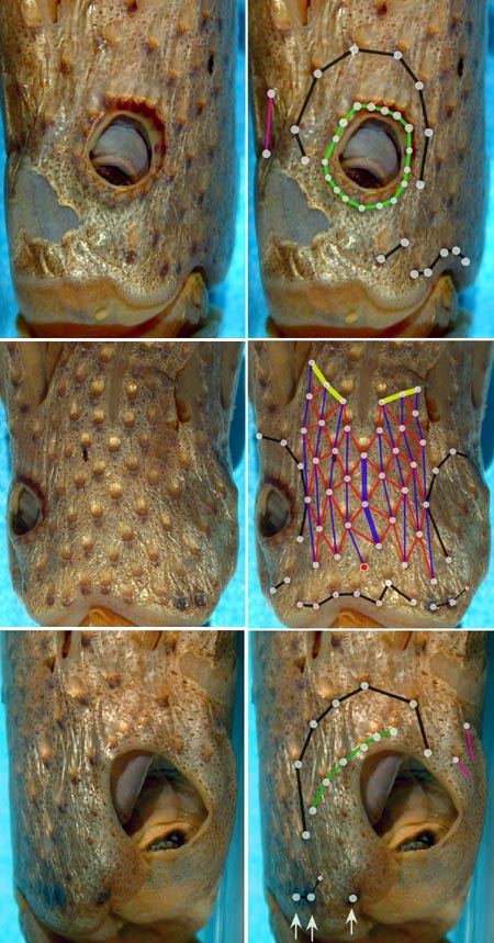
Figure. Right, ventral and left views of the head showing photophores of H. bonnellii, NMNH specimen. Left - Unaltered photographs. Right - Same photographs but with the photogenetic region of each photophore indicated by a dot and with dots in each photophore series connected by a color-coded line
Click on an image to view larger version & data in a new window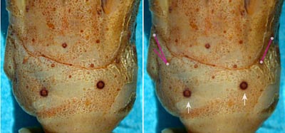
Figure. Paired dorsal views of the head of H. bonnellii, NMNH WH70. Left - Unaltered photograph. Right - Same photographs but with the photogenetic region of each compound photophore indicated by a white dot and with dots in the Right and Left Accessory Series of photophores connected by a violet line. Arrows point to unusually large simple-photophores. Photographs by R. Young.
- Photophores: Simple photophores (round, black dots):
- Arrangement not investigated.
- Two large, simple photophores on dorsal surface of head (see photograph below, white arrows).
- Small, simple photophores not easy to recognize and may be variable.
- Peculiar photophores
- 3 large, round, dark photophores on left posterior margin of ventral surface of head always present in squid larger than 18 mm ML (Voss, et al., 1998). The medial two appear to be compound photophores and, therefore, have been included in the Left Basal Series (see lower right photograph in above paired photographs).
Click on an image to view larger version & data in a new window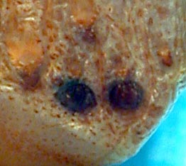
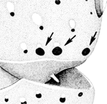
Figure. Ventral view of the left posteroventral region of the head of H. bonnellii. Left - Two large, dark medial photophores of the Left Basal Series. Photograph by R. Young. Right - Slightly more lateral view shows all three large, dark photophores, 42 mm ML, female, 42° 55'N, 60° 43'W. Drawing from Voss, et al., 1998.
- Mantle photophores
- Compound photophores of uniform large size on anterior half of ventral mantle.
Click on an image to view larger version & data in a new window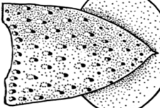
Figure. Ventral view of mantle of H. bonnellii, 21 mm ML, male, 43° 39'N, 24° 04'W. Drawing exracted from Fig. 35a of Voss, 1969.
- Arm photophores
- Arms IV with 3 longitudinal series on arm base.
- Single, elongate, dark, simple photophore on ends of all arms but much smaller on arms IV. The arm IV photophores do not appear until late juvenile or early subadult stages and may not appear in early maturing males in the southern half of the species range.
Click on an image to view larger version & data in a new window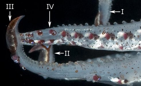
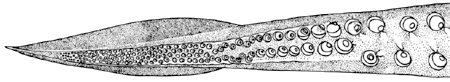
Figure. Top - Various views of the arms of a live H. bonnellii. Roman numerals refer to arm number. © . Bottom - Oral view of arm I tip of H. bonnellii, male, 21 mm ML, showsing the large aboral simple photophore, same as specimen listed on top. Drawing from Voss, 1969 (Fig. 35d).
- Tentacle
- Suckers of manus in 6 irregular series; median two series with suckers twice the diameter of ventral marginal suckers. Click on an image to view larger version & data in a new window
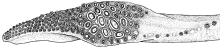
Figure. Oral view of the tentacular club of H. bonnellii, male 21 mm ML. Drawing from Voss, 1969 (Fig. 35b).
- Suckers of manus in 6 irregular series; median two series with suckers twice the diameter of ventral marginal suckers.
- Arms
- Length130-300% of ML.
- Length130-300% of ML.
- Sucker dentition
- Suckers of the club manus with numerous sharp teeth around entire margins.
- Arm sucker rings with 2-7 broad, blunt teeth on distal or distal and lateral margins.
- Web and buccal crown
- Inner web between arms I, II and III 50-60% length (mesured at midline of web) of longest arms.
- Buccal crown with 6 supports.
Click on an image to view larger version & data in a new window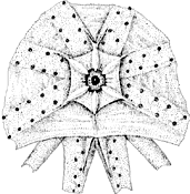
Figure. Oral view of buccal crown of H. bonnellii, male, 21 mm ML. Drawing from Voss, 1969 (Fig. 35g).
- Funnel organ
- Dorsal pad with median ridge on each lateral arm.
- Dorsal pad with median ridge on each lateral arm.
- Spermatophores
- 1.4-6.1% of ML; sperm mass 10-34% of spermatophore length (SpL); cement body 19-68% of SpL; ejaculatory apparatus 20-39% if SpL with 1-3 loops; connective complex present. Click on an image to view larger version & data in a new window
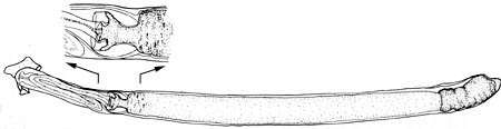
Figure. Spermatophore of H. bonnellii, 75 mm ML male, 38° 38'S, 51° 55'W, with enlargement of the area of the ejaculatory-apparatus. Drawings from Voss, et al. (1998). Compare this with the following spermatophore. Note the single loop of the ejaculatory apparatus, the short sperm mass and the constricted base of the ejaculatory apparatus/cement body complex.
Click on an image to view larger version & data in a new window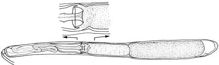
Figure. Spermatophore of H. bonnellii, 172 mm ML male, 20° 27'N, 21° 58'W, with enlargement of the area of the cement-body connective complex. Drawings from Voss, et al. (1998). Note the double loop of the ejaculatory apparatus, the long sperm mass and the lack of a constriction at the base of the ejaculatory apparatus/cement body complex. See "Distribution" for an interpretation of these differences.
- 1.4-6.1% of ML; sperm mass 10-34% of spermatophore length (SpL); cement body 19-68% of SpL; ejaculatory apparatus 20-39% if SpL with 1-3 loops; connective complex present.
- Hectocotylus
- Distal third of arms I in mature males modified. Each arm with two widely spaced series of small suckers of uniform size set on enlarged pedestals
Comments
With the exception of the analysis of the head photophores and the photographs, the above information is from Voss (1969) and Voss, et al. (1998).


