
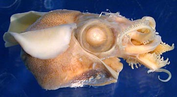

Figure. A. alveatus. Left - Lateral view, paratype no. 2, mature (?) male. Right - Ventrolateral view, holotype, mature male. Damaged ventral mantle resulted from a rusting pin used to orient the animal during drawning. Photographs by R. Young.
- Arms: Male
- Well-developed web between dorsal six arms.
- Well-developed lateral membrane joined to ventral side of arm IV well distal to basal suckers.
- Many suckers elongate with elongate, tubular inner rings, small aperatures and excentrically placed on long stalks.
- Arms I with biserial, normal suckers without obvious protective membranes or trabeculae.
- Arms II with approximately two series of normal suckers at base and two multiserial sets (clusters) of suckers distally separated by a short, bare region of the arm. Lateral suckers without protective membranes.
- Arms III with apparently normal suckers basally and a single greatly enlarged sucker (height 17% of DML) more distally with an elongate aperature. Membanes and trabeculae absent.
- Arms IV with elongate lateral suckers having elongate bases fused to protective membranes.
- Arms: Female
- Arms II with normal biserial suckers and thick lateral membranes distally sometimes forming separate lobes.
- Arms III with normal, approximate biseral suckers at base and bare distal half. Arm with irregular, lobular lateral membranes proximally and thick, transverse ridges distally.
- Arms IV with normal biserial suckers.
- Tentacles
- Tentacular clubs small, with tentacle-organ and larger proximal suckers (0.08 mm vs 0.04 mm).
- Head
- Eyes very large, 75% of lateral mantle length.
- Secondary ventral eyelid present.
- Occular pore apparently absent.
- Beaks not examined.
- Olfactory organ a small, round pad near dorsal opening of mantle.
- Funnel
- Funnel with well-developed lateral funnel adductor muscles.
- Funnel without a pore between medial adductor muscles.
- Mantle
- Dorsal mantle broadly fused to head between posterolateral corners of eyes and at the level of the anterior fin attachment.
- Mantle adductor weak.
- Anterior ventral mantle margin may be strongly indented.
- Photophores
- Large visceral photophore present with double lateral papillae.
- Gladius
- Not searched for, presumably absent.
- Measurements
*Measured to midpoint between eyes.Holotype Paratype no. 1 Paratype no. 2 Paratype no. 3 Sex Mature male Mature male Mature (?) Male Immature female Dorsal mantle length* 14 14 15 14 Ventral mantle length 16 18 18 17 Lateral mantle length 10 11 13
11
Mantle width 12 11 14 11 Head width 11 13 16 13 Eye diameter 8 7
9 7 Arm I length 6 6 7 Damaged
Arm II length 19 18 18 4 Arm III length 10 9 10 5 Arm IV length 13 13 14 7 Club length 3.3 4.0 Damaged Ca. 3.5
Fin length 11 13 14 13 Fin width 9 9 10 8

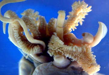
Figure. Oral view of arm crown of A. alveatus. Left - Holotype (reddish discoloration between the base of arms IV is from a rusting pin used to orient the animal during drawing. Right - Paratype no. 1.
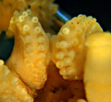
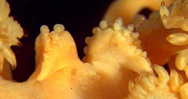
Figure. Arms I of A. alveatus, paratype no. 1. Left - Oral view of basal regions. Right - Dorsal-oral view of tips.

Figure. Oral view of the right arm II of A. alveatus, holotype. Drawing by Carolyn Gast, enlarged from illustration on species page.
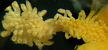

Figure. Two views of Arm II of A. alveatus. Left - Oral view of mid-region of arm with proximal set of suckers on the right separated from distal set on the left by a bare region of the arm, paratype no.1. The proximal set has smaller lateral suckers. Right - Oral view of the base of the arm showing approximate biserial arrangement of normal suckers giving way distally to many series of elongate suckers, holotype.

Figure. Aboral view of tip of Arm II of A. alveatus, holotype. Note the excentric attachments of the lateral suckers to the stalks.
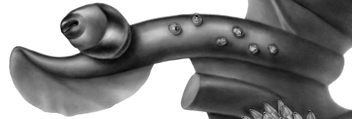
Figure. Oral view of the left arm IIII of A. alveatus, holotype. Drawing by Carolyn Gast, enlarged from illustration on species page.
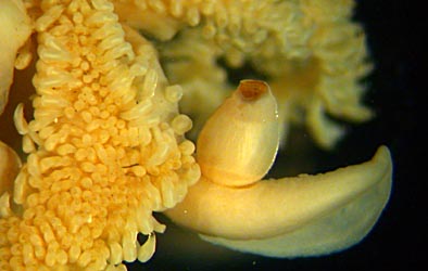
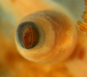
Figure. Two views of arm III of A. alveatus. Left - Ventral view showing some basal suckers, the enlarged, central sucker and the aboral flap (overlapping this arm on the left in the photograph is an oral view of a portion of arm IV), paratype no.1. Right - Oral view of the large arm III sucker showing the elongate aperature of its inner ring, holotype.
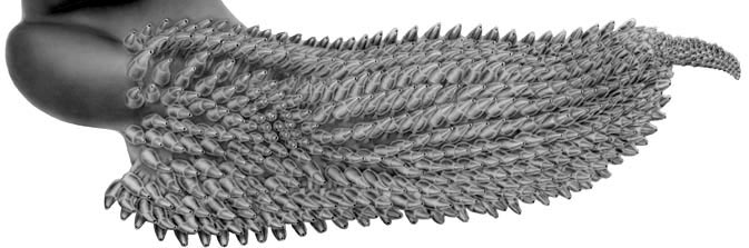
Figure. Oral view of the right arm IV of A. alveatus, holotype. Drawing by Carolyn Gast, enlarged from illustration on species page.

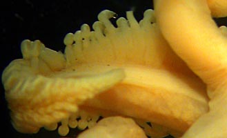
Figure. Two views of arm IV of A. alveatus. Left - Oral view of the tip of arm IV showing normal suckers, holotype. Right - Aboral view of dorsal region of the arm base showing joining of elongate stalks by a membrane that enables the broad lateral expansion of the arm, paratype no. 1.
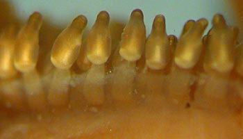
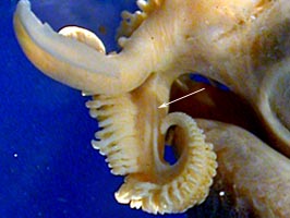
Figure. Two views of arm IV of A. alveatus. Left - Side view of the midarm showing a distinct membrane between stalks and the eccentric attachment of suckers to stalks, holotype. Right - Side view of arms III and IV showing the lateral membrane of arm IV (arrow) attaching well distal to the base of arm III, paratype no. 1.
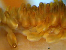
Figure. Various views of arm IV of A. alveatus. Side view of arm IV suckers, allowing view beyond lateral sucker series showing the near-tubular shape of the sucker inner rings and shorter stalks, holotype. *An oral view of the midregion of arm IV is seen in the photograph of the ventral view of arm III above.
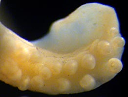
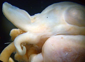
Figure. Arms of female A. alveatus, paratype no. 3. Left - Oral view of arm II showing biserial arrangement of suckers and aboral flap. Right - Lateral view of arms III and IV showing broad aboral flap on arm III and the broad connection between the bases of arms III and IV by the lateral membrane of arm IV which thereby forms the outer web for the tentacle pocket.
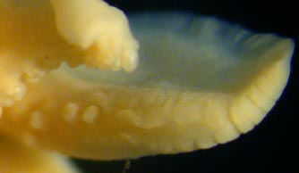
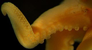
Figure. Arms of female A. alveatus, paratype no. 3. Left - Oral view of arm III. Arm III shows approximate biserial arrangement of suckers at arm base; distal arm shows lateral lobes giving way to transverse grooves. Right - Oral and lateral views of different regions of arm IV showing biserial arrangement of suckers.
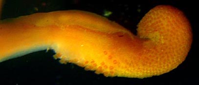
Figure. Oral view of left tentacular club of A. alveatus, holotype. Note large dorsal tentacle-organ and large proximal suckers. Some large suckers appear to have been lost during capture.
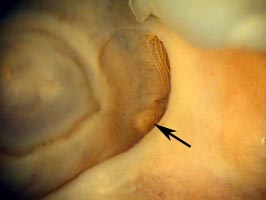
Figure. Lateral view of the olfactory organ (arrow) of A. alveatus, female, paratype no. 3.

Figure. Ventral view of anterior mantle margin A. alveatus showing strong indentation, holotpe.
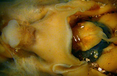
Figure. Ventral view of the visceral photophore of A. alveatus, female, paratype no. 3. Note the two papillae (arrows) on either side of the photophore which have terminal pores that presumably lead into the lumen of the photophore. Debris in the photograph is sand grains.


