Drawings below are first made from photographs. The drawings should then be compared, under a microscope, with the preserved squid from which the photographs were made. This greatly reduces the errors that can occur in interpreting the photophore distributions. Use caution, therefore, on this page in viewing the color and distribution of the dots (representing photophores) as their distributions have not yet been confirmed by comparing the drawings against the actual specimen under the microscope.

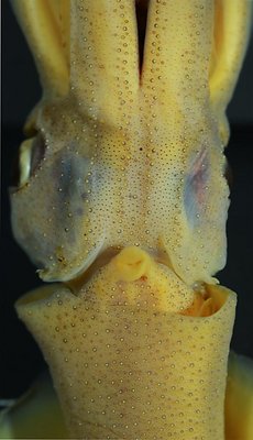
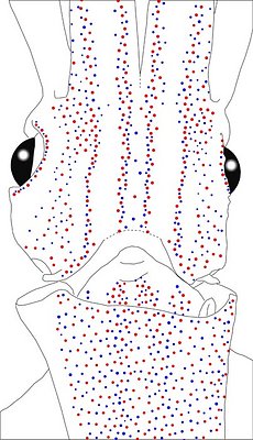
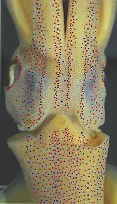
Figure. Ventral views of the integumental photophores of A. gilchristi, female, 32 mm ML, South Atlantic. Left - Photograph of the preserved squid. Middle - Outline drawing from photograph with all integumental photophores represented by colored dots. Right - Photograph with colored dots superimposed on integumental photophores. Red dots - Complex photophores. Blue dots - Non-complex photophores. Images by R. Young.
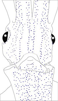
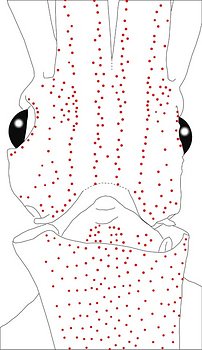
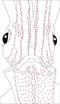
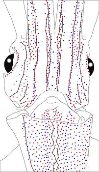
Figure. Same squid, same view as above. Left - Drawing showing only the non-complex (blue) photophores. Left middle - Drawing showing only the complex (red) photophores. Right middle - Same as previous drawing with lines connecting red photophores to aid comparisons of photophore patterns between species. Right - Drawing showing all photophores and lines showing red photophore patterns. Images by R. Young.
Figure. Ventral view of the mantle of the same squid (32 mm ML). Note that the Median Mantle Sector is not easily identified. Photograph by R. Young.
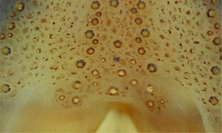
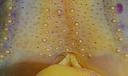
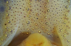
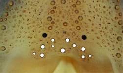
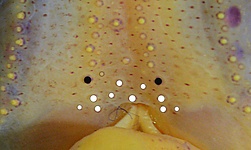
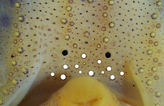
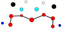
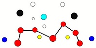
Table. Ventral view of the funnel-groove region of A. gilchristi. Top tier - Funnel groove showing photophores of funnel groove and surrounding tissue. Middle tier - In the photograph, white dots placed over the "white" photophores of the funnel groove. Black dots placed over the two most posteromedial "red" photophores of the ventral head to mark the anterolateral edges of the funnel groove. Bottom tier - Same pattern of dots as in photographs but dots are colored. Colored dots, lines - Aid in comparison of patterns between species. Open dots - Represent photophores that are either of uncertain status as "white" photophores or have variable presence. Images by R. Young. Comparison of white photophore patterns for all Abraliopsis species can be found here.
| Mature (?) male, 32 mm ML | Mature (?) female, 25 mm ML | Mature (?) female, 32 mm ML |
|---|---|---|
 |  |  |
 |  |  |
 |  |  |
Comments 1:
The photophore arrangements of Abraliopsis (Micrabralia) gilchristi are very different from all other members of the genus. In many ways the arrangements seem intermediate between those of the subgenus Abraliopsis, and those of Pfefferiteuthis. Red photophores are scattered on the mantle and in some areas of the head. The arrangement of head series is unique with 4 series (at least) and with each Medial Head Series being very irregular or, perhaps, better considered as a double series.
Comments 2: Summary of photophore distributions.(photophore definitions found here) ****
ARMS:
Medial Arm IV Series - Continuous; reaches about 2/3 of arm length.
Central Arm Sector - Few blue photophores at base near margins (assuming a double red series at arm base).
Lateral Arm IV Series - Slightly discontinuous (?), apparently with 1 or 2 small breaks in midarm; reaches arm tip.
Lateral Membrane Series of arms IV - Reaches at least 3/4 of arm length.
Arm III series - Continuous, reaches at least 3/4 of arm length.
HEAD - Unique four photophore series on central-ventral head with:
Median Head Series - Absent.
Median Head Sector - Few red and blue, scattered photophores.
Medial Head Series - We consider this series to be a double series with a few red photophores lying between the two branches of the double series.
Lateral Head Sector - A few, peripheral blue photophores anteriorly, otherwise, photophores absent.
Window Sector - Scattered red and blue photophores at anterior and posterior ends.
1st Lateral Window Series -Two segments broadly separated by single series of blue photophores: At 32 mm ML, posterior segment with 4 red photophores; anterior segment with 3 reds that join 4 reds of the Lateral Membrane Series of arm IV.
2nd Lateral Window Series - Continuous; 10-11 red photophores at 32 mm ML.
Eyelid Series - Encircles eye opening with red photophores only on ventral half. Posterior Eyelid Series with one large blue photophores displaced slightly posteriorly from eyelid series. Formula: RbBbB or RbbBB (depending on viewpoint).
Occipital Series - RbbRbR
Lateral Funnel-groove Series - Two parts: posterior part - 2 reds; anterior part - 4 reds (32 mm ML). Could be interpreted as 3 reds posterior and 3 anterior.
Olfactory Series - 2 red photophores at 32 mm ML. Posterior member just posteroventral to first photophore (= most posterior) of 1st Lateral Window Series and nearest red photophore to olfactory organ on 2nd occipital fold. Anterior member anteroventrally to posterior member. This series also present in Abraliopsis sp. Z.
White patch (funnel groove) - Moderate pattern (red, green, blue + yellow with size).
FUNNEL:
Medial patch - Scattered red photophores.
Lateral patch - Scattered red photophores.
MANTLE - Scattered photophore pattern with:
Median Sector - Often not easily recognized.
Medial Mantle Series - Narrow and very irregular
Lateral Mantle Sectors - 1st Sector with greater concentration of photophores than 2nd Sector.
Lateral and Mantle-angle Series - Lateral Series not easily recognized. Mantle-angle Series more typical.


