The "histioteuthid-club" of squid in the Histioteuthidae and Psychroteuthidae differs greatly from all other decapodiform tentacular clubs. Here we compare the structure of its manus with that of a "basic oegopsid club" of Ommastrephes bartramii.
1. The most apparent difference between the two clubs is the appearance of an accessory muscular lobe in the histioteuthid club that virtually doubles the cross-sectional size of the manus.
2. The muscular stalks of all the ommastrephid suckers are constricted where they attach to the suckers while the large histioteuthid suckers have little or no constriction.
3. The sucker stalks of the histioteuthid club originate on the surface of the club core (suckers 3 and 5), while some of the intermediate to small suckers (suckers 4 and 6) originate on the surface of the accessory lobe. The origin of the small suckers near the ventral margin (suckers 1, 2) could not be clearly determined. In the ommastrephid club, all sucker stalks and muscular trabeculae originate on the club core.
4. Suckers on the histioteuthid manus form irregular series and these number more than four, while the ommastrephid manus has suckers in four regular series. Clubs with more that four sucker series, however, probably arose numerous times in the Oegopsida.

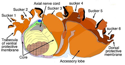

Figure. Left - The "histioteuthid-club" of Histioteuthis reversa. Right - A basic oegopsid club of Ommastrephes bartramii. Dots indicate longitudinal muscles, hash marks indicate mostly transverse muscles; curved lines indicate complex muscles. Note that in the left image, the stalk of sucker 5 originates on connective tissue covering the club core, extends through the accessory lobe to a large sucker located or the oral surface of this lobe. The accessory lobe as a peculiar shape with a looping extension that attaches firmly to the club core only at the aboral end of the core. The stalks of the large suckers (e.g., sucker 5) are mostly free (i.e. in contact with sea water) for their full length except for fusion along one side. The large keel of the ommastrephid manus is not seen on the histioteuthid-club as the latter keel is present on the dactylus but terminates proximally at the edge of the manus. Drawings, reconstructions from serial, histological sections, by R. Young.
We have not re-constructed histological sections of the psychroteuthid club but a quick glance at a single section through the manus (below) shows the great similarity to the club of Histioteuthis reversa (above) and particularly the unusual accessory lobe. The photograph below also show how complex the musculature is.
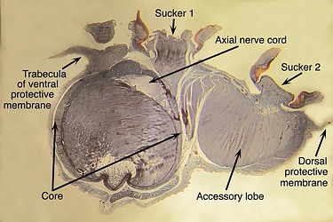
Figure. The "histioteuthid-club" of Psychroteuthis glacialis. Histological section through the manus that is lightly stained with methylene blue dye. Photographs by R. Young.
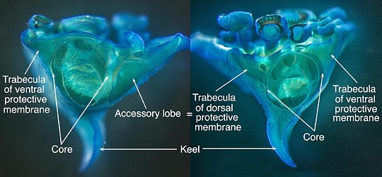
Figure. Fragment of a club of Stigmatoteuthis hoylei cut at the transition between the manus and dactylus and lightly stained with methylene blue dye. Thickness of the fragment is about 3-4 suckers deep. Left - Cut viewed from the proximal (= manus) side of the fragment. Right - Cut viewed from the distal (dactylus) side of the fragment. Photographs by R. Young.
[A cut through the manus of the histioteuthid S. hoylei (not shown) looks just like the similarily placed cut of H. reversa above.]
A very thick segment of the club, cut from the club of S. hoylei (presumably H. reversa would be similar) from near the beginning of the dactylus, gives a different perspective on the club structure. In the cut nearest the manus (Left image), the dorsal trabecula of the protective membrane is an extension of the reduced accessory lobe. The cut nearest the dactylus (Right image) appears to be a rather typical oegopsid club with similar trabeculae on either side (compare with the ommastrephid drawing). The accessory lobe, therefore, appears to result from a fusion and elaboration of the trabeculae of the protective membrane of the dorsal side of the manus. In both dorsal and ventral membranes, individual trabeculae of clubs of large squid cannot be detected; however, in a young H. reversa individual trabeculae, adjacent to one another, can still be recognized (Naef, 1921-23, Fig. 178).
When one looks at the width of the oral face of a histioteuthid-club relative to the width of its tentacle stalk, it doesn't seem to be much different than a similar comparison made with an O. bartramii club and stalk. This suggests that the core of a histioteuthid club is relative small and that the function the the accesssory lobe and its associated modifications is more than just to increase the width of the club. In cross-section, the accessory lobe is loosely attached to the club core (this is unusual for a "trabecula"), except at the aboral end of the core; in contrast, the stalks of the larger club suckers are strongly attached at their base to the core but otherwise are mostly free (i.e., bathed in sea water) except along their distal side. We assume that this arrangement somehow allows the histioteuthid-club a greater range of flexibility in grabbing objects (mostly prey) than does the ommastrephid club.


